Professional & Compassionate Eye Care Services & Exams
Dr. Markis-Meyer and associate doctors provide comprehensive eye examinations for all patients. They will review your medical and ocular history, check your eye alignment, movement, peripheral vision and pupil health. Our doctors will also check your visual acuities and measure the glasses you are wearing (if any).
The main portion of the eye exam includes a refraction, which checks your focusing ability at both near and far distances and determines your prescription. We have state-of-the-art eye examination equipment to provide you with the most technologically advanced measurement.
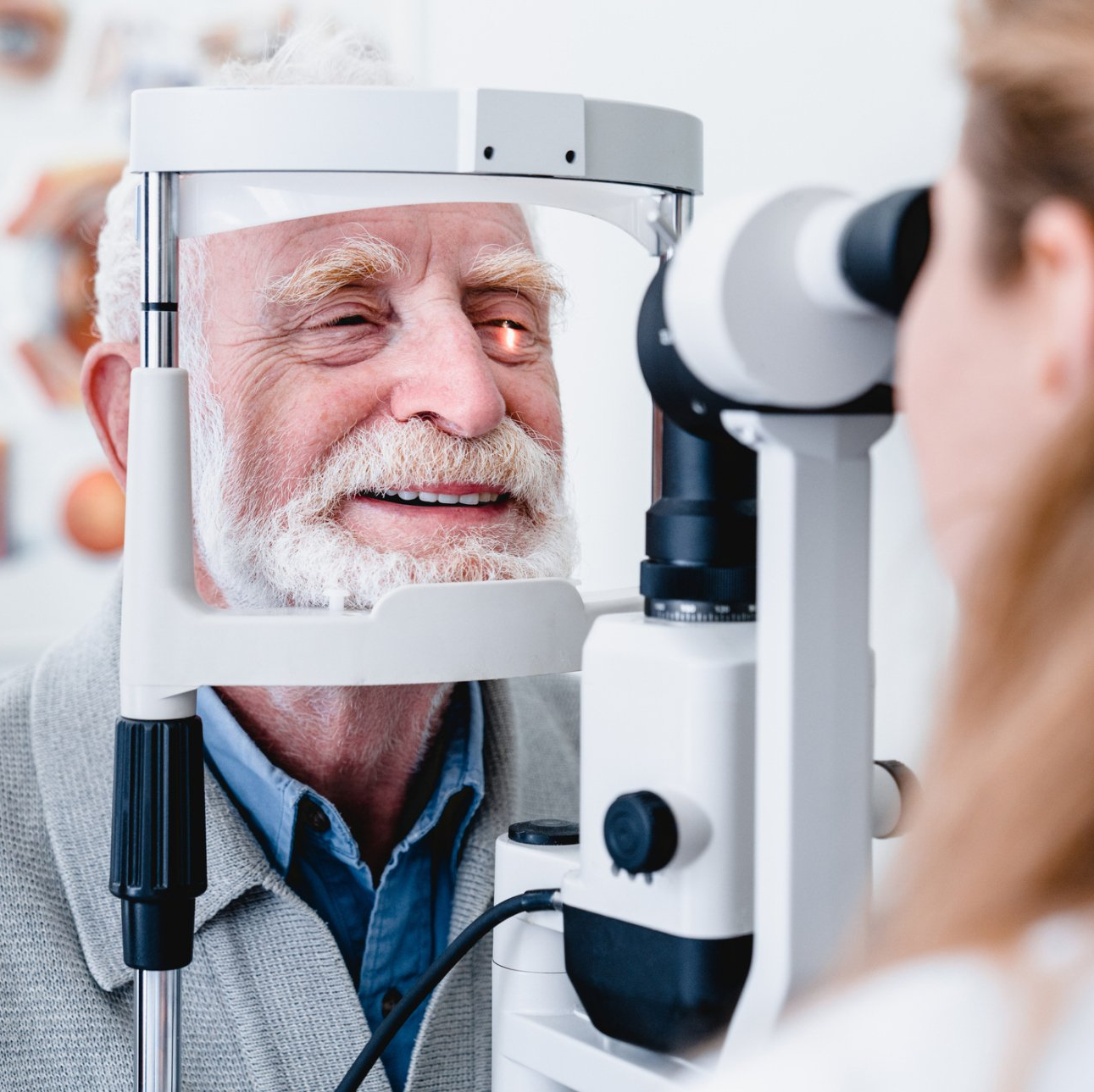
After discussing your options regarding glasses, our doctors will check the health of your eyes. This includes testing for glaucoma and examining the structures inside the eye with a special microscope called a slit lamp. The best way to inspect the internal structures of the eye is by either dilating the pupil where the doctors can get a clear view of the lens (to check for cataracts) and the retina (to check for various retinal diseases such as macular degeneration) or the optomap wide-field retinal images. This internal examination of the eye should be done every year, or more often if you have diabetes or another condition that needs to be closely monitored. Pupil dilation will make you extremely light sensitive and make your near vision blurry for 3 to 4 hours. If it is not convenient, another time can be scheduled for you to be dilated or you can opt to have retinal images taken in lieu of dilation.
Recommendations for Eye Exams
All children should have their eyes checked by age 5. If there is any family history of childhood vision problems, or if the child has developed signs or symptoms of a vision problem, they should be checked earlier.
Between the ages of 5 and 20, you should have an eye exam every 2 years.
- age 20 and over every year
If you wear contact lenses, you will need to be checked every year, regardless of age.
You should be seen if you notice a change in your vision or if you experience pain, flashes of light, new floaters, tearing, or if you sustain injury to the eye.
Optomap® Retinal Exam
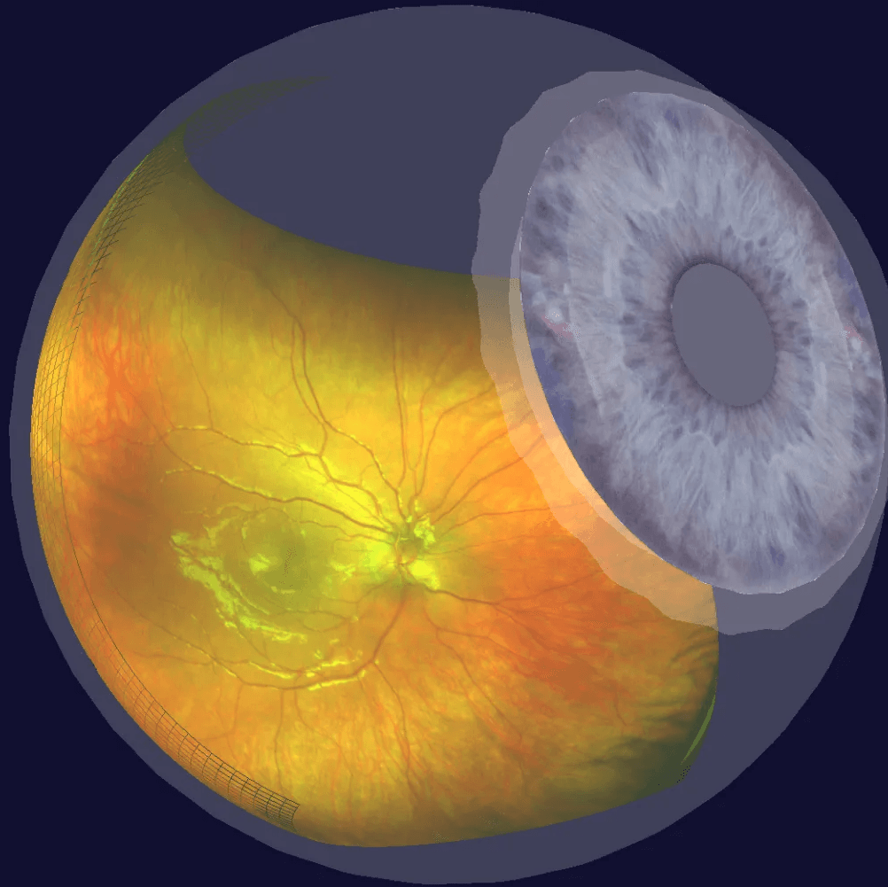
At Grass Valley Eye Care, we pride ourselves on providing our patients with the best possible standard of care. Because of this, we now perform the optomap® Retinal Exam with all of our patients. This non-invasive procedure allows your doctor to see a much broader and more detailed view of the retina than is possible with conventional methods. When reviewed, the scan becomes a permanent part of your medical file, enabling your doctor to make important comparisons should potential vision-threatening conditions show themselves at a future time. Our doctors will review the images and share the 3D view of the inside of your eye during your exam. We strongly believe that the Optomap® Retinal Exam is an essential part of your comprehensive eye exam and prescribes it for all patients once per year, and it is part of your pretest workup.
**FOR MOST SITUATIONS, THIS TEST DOES NOT REQUIRE DILATION**
As part of your pre-test workup, we will capture optomap® images for review with your doctor during your examination today. The doctor needs to view the retina. This advanced technology is convenient, informative, and a significant option recommended at Grass Valley Eyecare Optometric, Inc. The price is $39.00. Some photos may be billable to your medical insurance. Any questions you have about the optomap® Retinal Exam can be directed to Dr. Markis-Meyer or Dr. Alexander when the images are reviewed during your examination.

